Oligohydramnios
Too Little Amniotic Fluid
![]()
![]()
Oligohydramnios |
 |
OligohydramniosContents • What Is Oligohydramnios? |
Return To Main Guide |
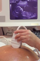
What Is Oligohydramnios?
It is a condition where too little amniotic fluid surrounds the fetus in the womb (image). Amniotic fluid cushions the baby and is a vital part of its life support system in aiding the development of its muscles, lungs and digestive system. About 8 percent of pregnant women will experience low amniotic fluid levels, although only 4 percent will be diagnosed with oligohydramnios. The majority of women with oligohydramnios still go on to have completely normal pregnancies although there is a slight risk of umbilical cord restriction if there is too little fluid for the baby to float around in. In most cases the condition is caused by a small puncture in the amniotic sac. In less common occurrences it can be an indication of a problem with the baby such as intrauterine growth restriction (IUGR). Oligohydramnios usually occurs in the latter part of the third trimester, although it can happen earlier. Women are at increased risk for the condition if they go past their due date as amniotic fluid levels decrease by nearly 50 percent after week 42. It causes complications in 12 percent of pregnant women past week 41. On the other side of the scale, if amniotic fluid levels are too high, this condition is medically termed polyhydramnios.
Leaking fluid from the vagina is one sign to watch out for. If you notice it, report it to your pregnancy team immediately. In most cases however, the mother experiences no symptoms. It is usually detected accidentally during routine prenatal pregnancy ultrasound scans. Warning signs are when the uterus measures smaller than usual, if the baby is not growing as fast as it should or if the mother is not gaining enough weight. Oligohydramnios is usually diagnosed by ultrasound scan. It can be discovered by chance during routine prenatal scanning or noted by a doctor checking for other conditions (antepartum surveillance). Using ultrasound the doctor measures the amount of amniotic fluid present. There are two ways to do this: maximum vertical pocket (MVP) and amniotic fluid index (AFI). MVP measures the deepest area or quadrant of the uterus for amniotic fluid levels. If the MPV score is less than 2cms then a diagnosis of oligohydramnios is given. AFI checks fluid levels in all 4 quadrants or pockets of the uterus. These amounts are added together and if the AFI score is less than 5cm then oligohydramnios is diagnosed. Both AFI and MVP are diagnostically accurate, although MVP may be better for assessing a mother expecting multiple births or where the pregnancy is still in its early stage. The doctor will also scan the fetus to rule out serious potential causes. For example, if the fetus' kidneys and bladder appear normal, the doctor can rule out cystic dysplasia and renal agenesis (both kidney disorders) as well as urethral obstruction. A Doppler ultrasound will be used to assess fetal growth if IUGR is suspected.
Ruptures Mothers Health Medications Placental Issues Birth Defects Over-Due What Complications Can It Cause? If oligohydramnios occurs in the first 6 months of pregnancy (which is less common), it is more likely to cause medical emergencies than if it occurs in the third trimester. If it happens in the first 2 trimesters it can result in: 1. Miscarriage: Baby dies in the womb before week 20. If it happens in the third trimester it can result in: 1. The fetus to grow at a slower rate: Intrauterine growth restriction. Management of oligohydramnios is based on the gestational age of the fetus (how far advanced the pregnancy is). Pre-Term Occasionally if the baby is still some weeks from its due date, amnioinfusion, a solution similar to amniotic fluid will be injected through the mother's belly to increase the fluid levels (by up to 30 percent). The doctor will normally monitor the procedure with an ultrasound scan. It will also help doctors visualize the baby’s physical anatomy better. However amnioinfusion is only a temporary measure because fluid levels can decline again within a week. The procedure can be repeated (known as serial amnioinfusion). Rehydration of the mother, either orally or by IV can also help to increase amniotic fluid levels. Bed rest or less physical activity may also be recommended. At-Term After-Term Vesicoamniotic Shunts If low amniotic fluid levels are caused by a blockage in the baby's urinary tract, a vesicoamniotic shunt may be used. A blockage can result in the baby's lungs or kidneys not developing properly, resulting in the necessity for dialysis or transplant after birth. During the procedure a needle is inserted through the mother's abdomen and into the womb. A flexible tube is moved down the needle and positioned between the bladder and the amniotic fluid. This allows fluid in the baby's bladder to drain into the amniotic cavity. The shunt is removed as soon as the baby is born and the procedure is carried out under local anesthetic. Children who undergo this treatment while in the womb very often go on to have the same quality of life as healthy children. However there is a 22–60 percent chance of the shunt becoming dislodged, so the procedure will need to be repeated. The earlier in pregnancy that oligohydramnios occurs, the poorer the outlook. Fetal mortality rates have been reported as high as 80 to 90 percent when oligohydramnios happens in the second trimester of pregnancy. Most deaths are the result of severe birth defects (congenital malformations). The prognosis for cases in the third trimester is very positive and most mothers, whose cases are properly managed, go on to deliver healthy normal babies. Other Pregnancy Complications • Bleeding during Pregnancy
|
| Related Articles
For more about care for pregnant women, see the following: • Prenatal care guide: For mom and baby. Return to Homepage: Womens Health Advice |
WOMENS HEALTH ADVICE: ABOUT PREGNANCY COMPLICATIONS |